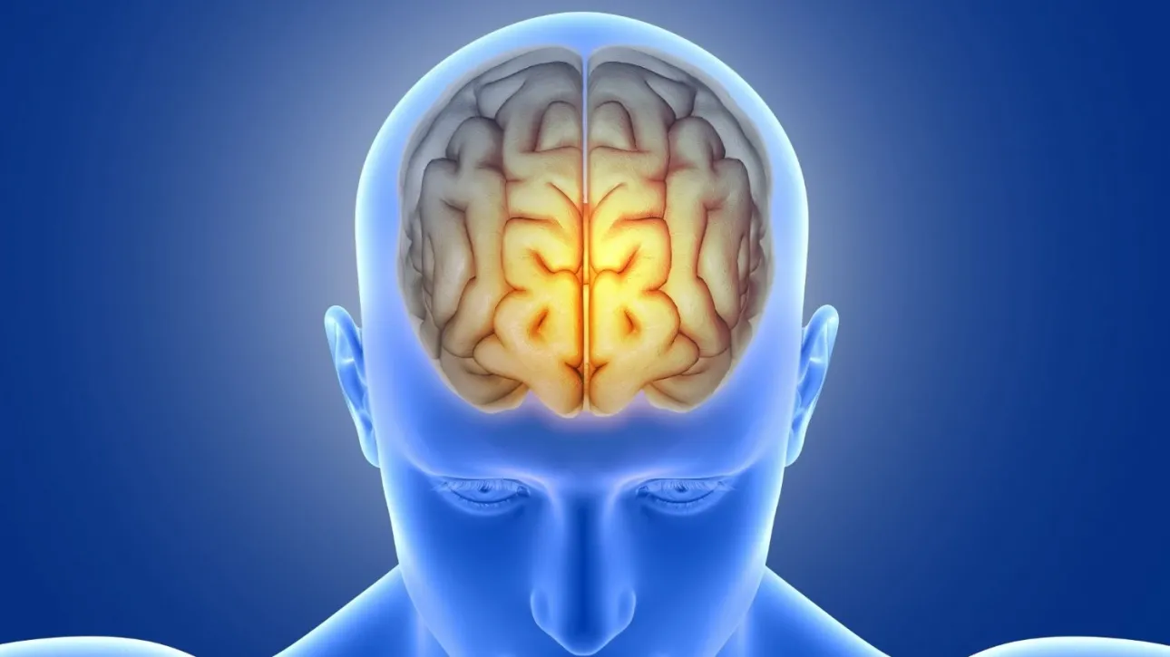
How does the brain remove its waste?
This task is carried out by the lymphatic drainage system, the study of which has led in recent years to significant advances in brain imaging.
A new study published in the scientific journal iScience reports the first direct evidence in humans of a previously unknown brain “cleaning hub”: the middle meningeal artery.
The research was conducted by scientists at the Medical University of South Carolina (MUSC), led by pathologist Onder Albayram. Through a collaboration with NASA, the team gained access to advanced real-time MRI tools originally designed to study how spaceflight affects fluid shifts in the brain. Using these tools, the researchers tracked the movement of cerebrospinal and interstitial fluid over a six-hour period in five healthy volunteers, according to SciTechDaily.
Rethinking the relationship between the brain and the immune system
Their observations showed that fluids moved along the middle meningeal artery slowly and passively, in a manner unlike arterial blood flow, which is faster and pulsatile. Instead, the pattern resembled lymphatic drainage.
“We observed a flow pattern that did not behave like blood moving through an artery. It was slower, more similar to drainage, indicating that this vessel is part of the brain’s cleaning system,” Albayram explained.
This discovery strengthens a broader reassessment of the scientific understanding of the relationship between the brain and the immune system. For decades, the prevailing view held that the meninges—the protective layers surrounding the brain and spinal cord—acted as a barrier, isolating the central nervous system from the lymphatic network. Over the past ten years, however, research by Albayram and others has shown that lymphatic vessels are embedded within the meninges and directly connected to the rest of the body.
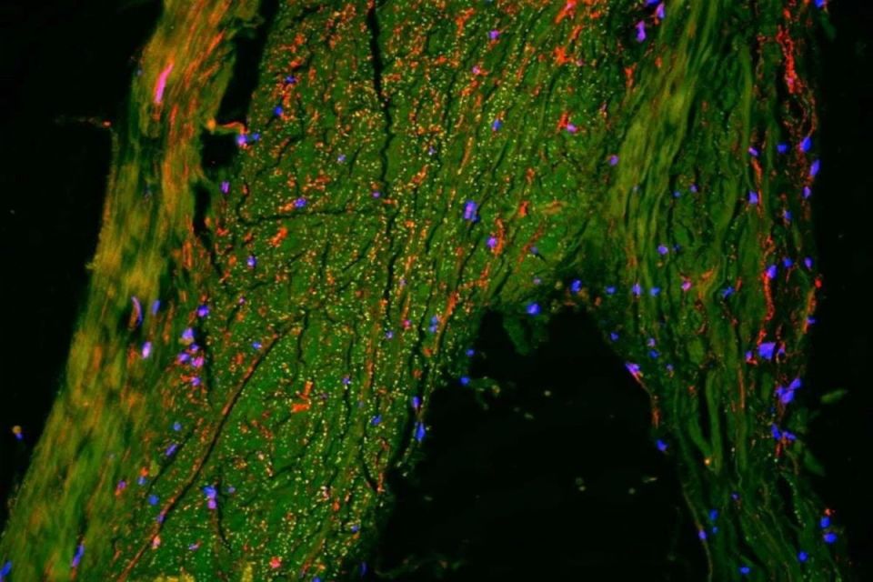
Microscope image showing lymphatic vessels (red) in the outer protective layer of the brain, known as the dura mater. These vessels form a rich and complex network that helps remove waste from the brain.
— Dr. Onder Albayram
To confirm the MRI findings, the researchers performed ultra–high-resolution imaging of postmortem human brain tissue. In collaboration with Cornell University, they used techniques that allow the simultaneous mapping of multiple different cell types. The analysis revealed cells characteristic of lymphatic vessels surrounding the middle meningeal artery, confirming that the slow fluid flow observed was not blood, but genuine lymphatic drainage.

Dr. Onder Albayram in his laboratory at the Medical University of South Carolina (MUSC).
Central to Albayram’s research philosophy is the study of healthy individuals before extending investigations to experimental models. Understanding how a “healthy” brain functions, he emphasizes, is a prerequisite for identifying early changes associated with injury, aging, neurodegenerative diseases, and psychiatric disorders.
“One of the major challenges in brain research is that we do not fully understand how a healthy brain functions and how it ages,” he notes. “Once we understand what is normal, we will be able to identify the early signs of disease and design better treatments.”