
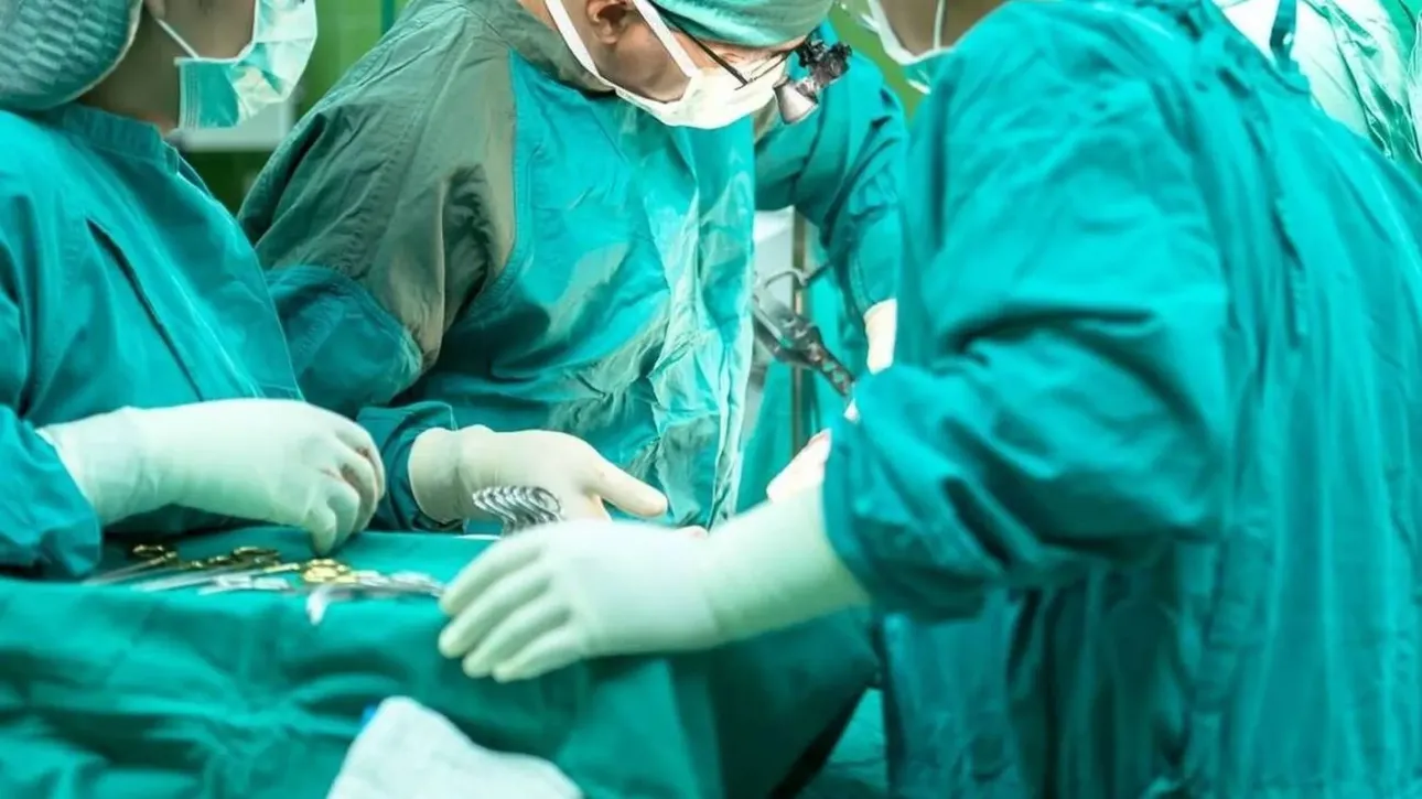
A new chapter in surgical medicine may have begun with the world’s first operation in which artificial intelligence was used in real time to support surgeons during the procedure. The operation was successfully completed, allowing doctors to remove a brain tumor from a patient who was at serious risk of losing his vision.
Reece Hibbert, a 48-year-old British father of two, underwent the pioneering procedure at University College London Hospital. According to the medical team, the specially designed AI system continuously analyzed live images from the surgical field and provided the surgeons with information about the location of critical blood vessels and nerves that were not directly visible.
This allowed the surgical team to approach the tumor more precisely and remove as much of it as possible while reducing the risk of damaging vital anatomical structures.
“From a patient safety perspective, I completely understand the benefit. There are no road signs inside our heads,” Hibbert said.
From a sudden seizure to a diagnosis
Until around 18 months ago, Hibbert was healthy and physically active. His life changed when he suddenly lost consciousness during a walk and began experiencing seizures in the street.
Brain imaging revealed an approximately 11 millimeter benign tumor in his pituitary gland, a small gland located at the base of the skull.
The pituitary gland produces hormones involved in several essential bodily functions, including growth, metabolism and the regulation of blood pressure and body temperature.
The tumor’s location presented a significant surgical challenge because it was close to the carotid arteries, major blood vessels supplying the brain, as well as the optic nerves responsible for vision.
As the tumor began pressing on Hibbert’s optic nerves, his field of vision became increasingly restricted.
Fatigue, dizziness and the fear of losing independence
Although surgery was not immediately necessary, his condition began affecting his everyday life. Hibbert started stumbling more frequently and experienced severe fatigue and dizziness.
He said the greatest impact was not necessarily physical but emotional and psychological.
“Although many people kindly offered to help, you don’t want to become a burden to other people,” he said.
When doctors offered him the opportunity to become the first patient to undergo the procedure using the new AI system, he did not hesitate.
“If patients aren’t willing to take part in research, how will doctors ever learn and how will medicine progress?” he said.
A procedure in a highly challenging anatomical region
The operation was performed through the nose. Surgeons inserted an endoscope—a thin camera mounted on a shaft and advanced it toward the base of the skull.
The tumor was located behind layers of bone and tough membranes, making access particularly challenging.
Finding the safest route to remove it is not straightforward because human anatomy varies from one patient to another.
The risks can be significant. Depending on the case, there is a 25%-50% chance that part of the tumor may remain, while the risk of damaging a major blood vessel is estimated at approximately 0.5%-2%.
AI acts as a “second pair of eyes”
This is where the new technology comes into play.
The AI system was displayed on a separate screen and continuously analyzed the live video feed from the operation. It tracked surgical instruments and identified areas where important blood vessels and nerves were likely to be located, while indicating regions where approaching the tumor was likely to be safer.
The technology works in a way broadly comparable to facial-recognition systems found in smartphones. Instead of recognizing faces, however, the system has been trained to identify critical anatomical structures, including those that may not be directly visible.
Researchers trained the model using hundreds of videos from pituitary tumor operations. They carefully mapped the location of blood vessels and nerves in each recording, creating the dataset needed to teach the system to recognize the most important structures.
Professor Hani Marcus, who was involved in the procedure, said the system has effectively been trained on more operations than a surgeon could observe throughout an entire career and can serve as an additional, experienced pair of eyes.
One of its most important features is its ability to analyze the surgical scene in real time, unlike several other experimental AI tools that are primarily used before or after surgery.
The surgeon remains in full control
Despite the technology’s capabilities, the team behind the procedure stresses that its purpose is not to replace surgeons.
The AI system is designed to provide support, while the final decisions and full control of the operation remain with the medical team.
The researchers have an even more ambitious long-term goal: developing a kind of “ChatGPT for surgeons”, a digital assistant capable of providing advice and information inside the operating room whenever doctors request it.
As Professor Marcus explained, such a system could effectively become “another expert in the room” one surgeons could consult for a second opinion, or ignore if they disagreed with its recommendation.
The procedure represents only an early step. But it demonstrates how artificial intelligence could move beyond analyzing medical data and into active, real-time support during surgery, potentially improving precision and safety for patients.